

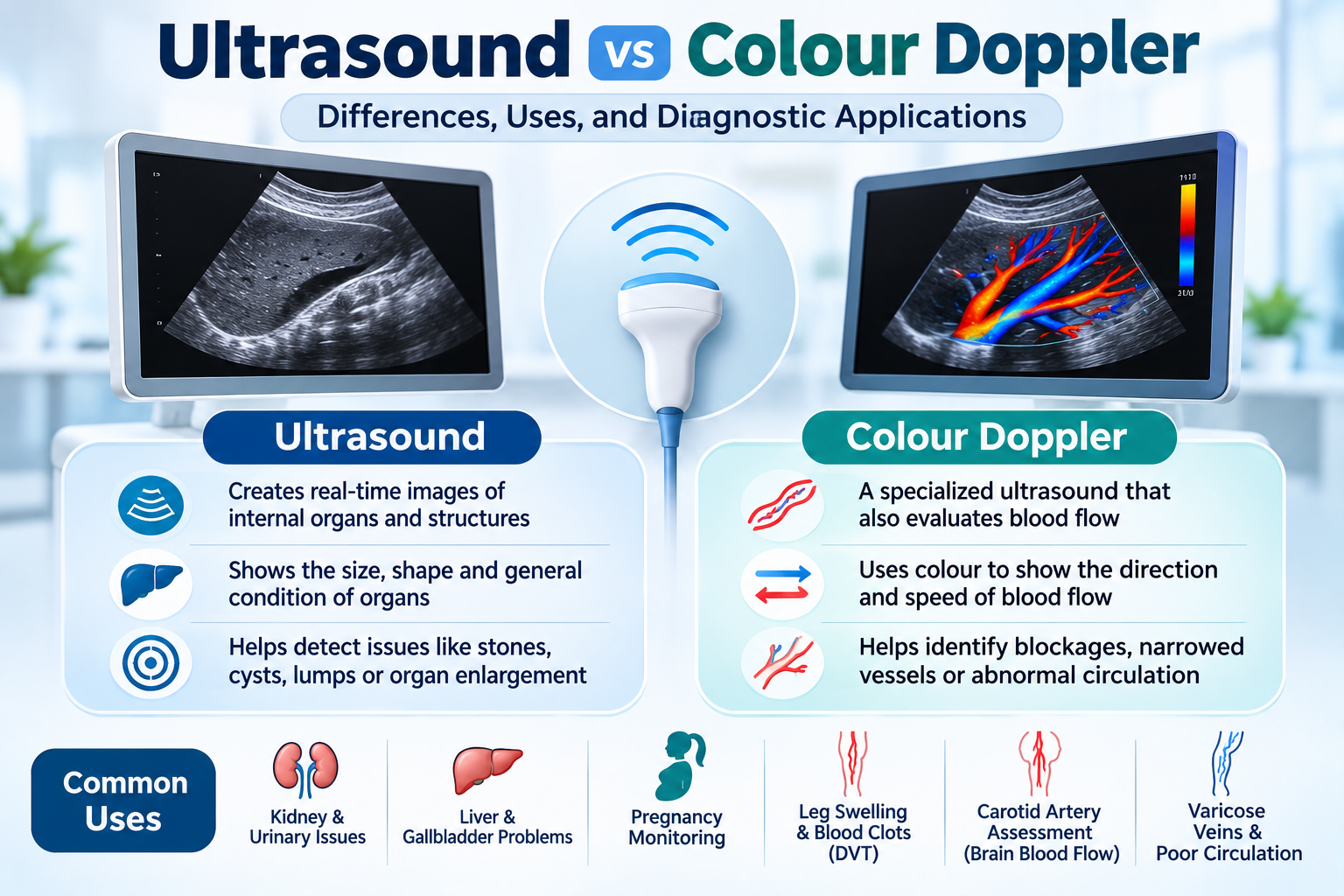
When a doctor recommends an ultrasound or Colour Doppler test, many patients wonder whether both procedures are the same. Although they use similar technology, these diagnostic tests serve different purposes.
An ultrasound primarily helps doctors examine the structure of internal organs, tissues, and other body parts. A Colour Doppler ultrasound, on the other hand, provides additional information about blood flow through arteries and veins.
Both are widely used diagnostic imaging techniques that help doctors investigate medical conditions without using ionizing radiation.
Understanding the difference between ultrasound and Colour Doppler can help you know why a particular test has been recommended and what information it can provide.
What Is an Ultrasound Test?
An ultrasound, also known as sonography, is a diagnostic imaging procedure that uses high-frequency sound waves to create images of structures inside the body.
During the examination, a healthcare professional moves a small device called a transducer over the skin. The transducer sends sound waves into the body and receives the echoes that return from internal structures. These signals are converted into images displayed on a monitor.
An ultrasound may be recommended to examine:
- Liver and gallbladder
- Kidneys and urinary bladder
- Pancreas and spleen
- Uterus and ovaries
- Prostate gland
- Thyroid gland
- Muscles, tendons, and soft tissues
- Developing baby during pregnancy
What Conditions Can an Ultrasound Help Detect?
Depending on the body part being examined, ultrasound can help identify:
- Gallstones
- Kidney stones
- Fatty liver changes
- Liver enlargement
- Ovarian cysts
- Uterine fibroids
- Prostate enlargement
- Fluid accumulation
- Certain soft tissue abnormalities
The findings help doctors decide whether further investigations or treatment are necessary.
What Is a Colour Doppler Test?
Colour Doppler is a specialized ultrasound technique that evaluates blood flow through blood vessels and certain parts of the heart.
Unlike conventional ultrasound, which primarily provides images of internal structures, Colour Doppler uses changes in sound-wave frequency to assess the movement of blood.
The information is displayed using colour overlays that represent the direction and relative velocity of blood flow.
Colour Doppler is commonly used to examine:
- Arteries supplying blood to different body parts
- Veins carrying blood back towards the heart
- Blood flow in the neck and carotid arteries
- Blood circulation in the arms and legs
- Blood vessels in the abdomen
- Blood flow in certain pregnancy-related examinations
What Conditions Can Colour Doppler Help Detect?
Depending on the type of examination, Colour Doppler may help identify:
- Deep vein thrombosis (DVT)
- Narrowing of arteries
- Reduced blood circulation
- Blood vessel blockages
- Varicose veins and venous reflux
- Abnormal blood flow patterns
- Certain vascular abnormalities
It is particularly useful when doctors suspect problems involving circulation or blood vessels.
Ultrasound vs Colour Doppler: Key Differences
Although both tests use ultrasound technology, their primary objectives are different.
| Feature | Conventional Ultrasound | Colour Doppler |
|---|---|---|
| Main purpose | Examines internal structures | Evaluates blood flow |
| Technology | High-frequency sound waves | Doppler ultrasound technology |
| Image appearance | Grey-scale images | Grey-scale images with colour flow overlays |
| Blood circulation | Not routinely assessed | Specifically evaluates blood flow |
| Common applications | Liver, kidneys, abdomen, pelvis | Arteries, veins and vascular circulation |
| Detects | Cysts, stones, organ changes and fluid | Reduced flow, abnormal flow and vascular problems |
Important: Colour Doppler is not a completely separate technology from ultrasound. It is an additional ultrasound technique that provides information about blood circulation.
When Do Doctors Recommend an Ultrasound?
A conventional ultrasound may be recommended when a doctor needs to examine internal organs or investigate symptoms such as:
1. Persistent Abdominal Pain
Ultrasound can help investigate possible gallstones, liver abnormalities, kidney problems, and other abdominal conditions.
2. Kidney or Urinary Problems
It may help identify kidney stones, urinary obstruction, bladder abnormalities, or changes in kidney size.
3. Gynaecological Concerns
Pelvic ultrasound can help evaluate ovarian cysts, uterine fibroids, and certain reproductive health conditions.
4. Pregnancy Monitoring
Obstetric ultrasound helps monitor fetal development, assess pregnancy progression, and evaluate selected pregnancy-related concerns.
5. Thyroid Abnormalities
Thyroid ultrasound can examine the size and structure of the thyroid gland and help evaluate thyroid nodules.
When Do Doctors Recommend Colour Doppler?
Colour Doppler is generally recommended when there is a need to evaluate blood circulation.
1. Leg Pain and Swelling
A venous Doppler examination may be recommended to investigate deep vein thrombosis or other venous conditions.
2. Suspected Artery Narrowing
Arterial Doppler can help assess blood flow and identify narrowing or reduced circulation in certain blood vessels.
3. Varicose Veins
Venous Doppler can evaluate blood flow through leg veins and identify abnormal backward flow caused by faulty valves.
4. Carotid Artery Assessment
Carotid Doppler helps examine blood flow through the arteries supplying the brain and can identify narrowing or plaque.
5. Pregnancy-Related Blood Flow Assessment
In selected pregnancies, obstetric Doppler examinations help evaluate blood flow through the umbilical artery and other relevant fetal or maternal vessels.
Is Colour Doppler More Advanced Than a Normal Ultrasound?
Colour Doppler is more specialized in terms of the information it provides, but it does not replace conventional ultrasound.
The two examinations answer different medical questions.
For example:
- If a doctor wants to examine the structure of your liver, an abdominal ultrasound may be appropriate.
- If a doctor wants to investigate blood circulation in your leg veins, a venous Colour Doppler may be recommended.
- If both organ structure and blood circulation need evaluation, the doctor may recommend a combined ultrasound and Doppler examination.
The choice depends on the symptoms, suspected condition, and clinical assessment.
How Are Ultrasound and Colour Doppler Tests Performed?
Both examinations are generally non-invasive and use similar equipment.
The procedure usually involves:
- Preparation: You may be asked to lie on an examination table.
- Gel application: A special ultrasound gel is applied to the area being examined.
- Image scanning: The sonographer moves the transducer over the skin to capture images.
- Doppler assessment: If required, Doppler settings are used to evaluate blood flow.
- Image review: The recorded images are reviewed and interpreted by an appropriately qualified medical professional.
The duration depends on the body part and complexity of the examination.
Most routine ultrasound and Doppler examinations take approximately 15–45 minutes, although some may take longer.
Do You Need to Prepare Before These Tests?
Preparation depends on the type of examination.
Before an Ultrasound
- Abdominal ultrasound may require fasting for a specified period.
- Pelvic ultrasound may require drinking water and having a full bladder.
- Pregnancy ultrasound preparation depends on the stage of pregnancy and scanning method.
- Other ultrasound examinations may require little or no preparation.
Before a Colour Doppler
Most Doppler examinations require minimal preparation. However, instructions can vary depending on the blood vessels being examined.
For example, an abdominal vascular Doppler may require fasting, while a leg venous Doppler usually does not.
Always confirm the preparation requirements with your diagnostic centre before the appointment.
Are Ultrasound and Colour Doppler Tests Safe?
Both procedures are generally considered safe when performed appropriately.
Their major advantages include:
- No ionizing radiation.
- Non-invasive examination.
- No surgical incisions.
- Real-time imaging.
- Generally painless procedures.
- Ability to assess different organs and blood vessels.
Some examinations may involve mild pressure from the transducer, but significant discomfort is uncommon.
Which Test Should You Choose?
There is no universal answer because ultrasound and Colour Doppler are designed to investigate different aspects of the body.
Your doctor may recommend:
- Ultrasound: When the main requirement is examining internal organs, tissues, or structures.
- Colour Doppler: When the main requirement is assessing blood flow through arteries or veins.
- Combined examination: When both structural and blood-flow information is necessary.
You should not select a diagnostic test solely based on personal preference. The appropriate investigation should be determined by your healthcare professional.
Final Thoughts
Ultrasound and Colour Doppler are valuable diagnostic imaging techniques that help doctors investigate different medical conditions.
While conventional ultrasound focuses primarily on examining internal organs and body structures, Colour Doppler provides additional information about blood circulation and vascular health.
Both tests have important roles in diagnosing medical conditions, monitoring certain diseases, and guiding further treatment decisions.
If your doctor has recommended an ultrasound or Colour Doppler examination, understanding the purpose of the test can help you feel more prepared and confident before your appointment.
At VR Diagnostics, patients can enquire about available ultrasound and Colour Doppler investigations and confirm the preparation instructions required for their examination.
Frequently Asked Questions
Is Colour Doppler the same as ultrasound?
Colour Doppler is a specialized ultrasound technique. It uses Doppler technology to evaluate blood flow, whereas conventional ultrasound primarily examines internal structures.
Which is better, ultrasound or Colour Doppler?
Neither is universally better. The appropriate test depends on what the doctor needs to investigate. Ultrasound examines organ structures, while Colour Doppler evaluates blood circulation.
Can Colour Doppler detect blood clots?
Venous Doppler ultrasound is commonly used to investigate blood clots, particularly deep vein thrombosis in the legs. The results must be interpreted by a qualified healthcare professional.
Does Colour Doppler require fasting?
Most routine vascular Doppler examinations do not require fasting. However, certain abdominal Doppler studies may require it. Always follow the instructions provided by your diagnostic centre.
How long does a Colour Doppler test take?
Many routine Colour Doppler examinations take approximately 20–45 minutes. The actual duration depends on the body area and complexity of the examination.
Is Colour Doppler safe during pregnancy?
Ultrasound-based Doppler examinations are generally considered safe when medically indicated and performed by trained professionals using appropriate settings. They should be used according to clinical recommendations.