
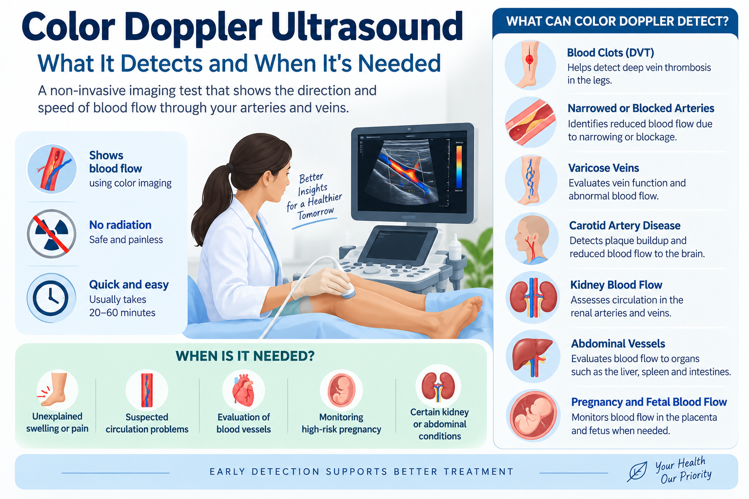
A Color Doppler ultrasound is a non-invasive imaging test that uses sound waves to examine blood flow through arteries and veins. Unlike a standard ultrasound, which mainly shows the structure of organs and tissues, Doppler imaging can help doctors assess the direction and speed of blood flow.
It is commonly used to investigate problems involving blood vessels, the heart, pregnancy, the abdomen, and the limbs. Since it does not use ionizing radiation, it is also a useful imaging option when Doppler assessment is clinically appropriate.
What Is a Color Doppler Ultrasound?
Color Doppler is an advanced form of ultrasound that combines conventional ultrasound imaging with Doppler technology.
The ultrasound probe sends sound waves into the body. When these waves reflect off moving blood cells, the system analyzes the change in frequency and displays information about blood movement.
The resulting images use colors to represent the direction and relative characteristics of blood flow, helping doctors identify areas where circulation may be abnormal.
What Can Color Doppler Detect?
Depending on the body part being examined, Color Doppler can help evaluate several conditions.
1. Blood Clots in the Legs
A venous Doppler ultrasound is commonly used when a doctor suspects deep vein thrombosis (DVT).
Possible symptoms include:
- Swelling in one leg
- Leg pain or tenderness
- Warmth
- Redness or discoloration
A Doppler examination can assess blood flow through the veins and help identify abnormalities that may suggest a clot.
2. Narrowed or Blocked Arteries
Doppler imaging can help assess blood flow through arteries and identify areas where circulation may be reduced because of narrowing or blockage.
This may be relevant in people experiencing:
- Leg pain while walking
- Cold feet or hands
- Numbness
- Slow-healing wounds
- Changes in skin color
3. Varicose Veins
Color Doppler can evaluate the functioning of veins and their valves.
It can help determine whether blood is flowing backward through incompetent valves, which may contribute to varicose veins and chronic venous insufficiency.
4. Carotid Artery Disease
A carotid Doppler examines the blood vessels in the neck that supply blood to the brain.
It can help identify:
- Plaque buildup
- Narrowing of the carotid arteries
- Abnormal blood flow
This examination may be recommended for selected patients with cardiovascular risk factors or symptoms suggestive of reduced blood flow to the brain.
5. Kidney Blood Flow
A renal Doppler examination can assess blood flow through the renal arteries and veins.
Doctors may recommend it when investigating certain kidney or blood-pressure-related problems.
6. Abdominal Blood Vessels
Doppler ultrasound can also assess blood flow through vessels supplying abdominal organs such as the liver, kidneys, and spleen.
It may be included as part of an ultrasound examination when vascular problems are suspected.
7. Pregnancy and Fetal Blood Flow
Obstetric Doppler examinations can assess blood flow through vessels associated with the placenta and fetus.
Depending on the pregnancy and clinical situation, doctors may use Doppler assessment to monitor circulation when there are concerns about:
- Fetal growth
- Placental function
- Maternal hypertension
- Certain high-risk pregnancies
The specific Doppler measurements performed depend on the doctor’s clinical assessment.
When Is Color Doppler Recommended?
A doctor may recommend a Color Doppler ultrasound when symptoms or other test results suggest a possible circulation problem.
Common reasons include:
- Unexplained limb swelling
- Leg pain or heaviness
- Suspected blood clot
- Varicose veins
- Poor circulation
- Carotid artery evaluation
- Certain kidney or abdominal vascular problems
- Monitoring blood flow during selected pregnancies
- Evaluation of vascular abnormalities
It is important to remember that not every swelling, pain, or circulation-related symptom requires a Doppler scan. The appropriate examination depends on the suspected condition.
Color Doppler vs Regular Ultrasound
Although both tests use ultrasound technology, they provide different information.
| Feature | Regular Ultrasound | Color Doppler |
|---|---|---|
| Shows organ structure | Yes | Yes |
| Evaluates blood flow | Limited | Yes |
| Shows direction of blood flow | No | Yes |
| Helps assess vascular narrowing | Limited | Yes |
| Uses ionizing radiation | No | No |
| Commonly used for blood vessels | Sometimes | Frequently |
In many cases, Color Doppler is performed alongside conventional ultrasound rather than replacing it.
How Should You Prepare for a Color Doppler?
Preparation depends on the area being examined.
For some abdominal examinations, you may be asked to avoid eating for several hours before the test. A full bladder may sometimes be required for certain pelvic examinations.
For Doppler studies of the legs, neck, or arms, special preparation is generally minimal.
Always follow the preparation instructions provided by your diagnostic centre or healthcare professional.
What Happens During the Test?
A Color Doppler ultrasound is generally straightforward.
During the examination:
- You will lie on an examination table.
- The sonographer will expose the area being examined.
- A small amount of ultrasound gel will be applied to the skin.
- The ultrasound probe will be moved over the area.
- Images and blood-flow information will be recorded.
- The examination usually takes between 20 and 60 minutes, depending on the body area and type of study.
You may feel mild pressure from the probe, particularly when examining certain blood vessels, but the procedure is generally painless.
Is Color Doppler Ultrasound Safe?
Yes. Ultrasound uses high-frequency sound waves rather than X-rays or other ionizing radiation.
It is widely used for diagnostic imaging, including during pregnancy when medically indicated.
However, ultrasound should still be performed for an appropriate clinical reason and interpreted by qualified professionals.
Does Color Doppler Require Contrast?
Usually, no.
Most Color Doppler examinations can assess blood flow without injecting a contrast agent. This is one of the advantages of ultrasound-based vascular assessment.
What Happens After the Scan?
The images are reviewed and interpreted by a qualified radiologist or appropriately trained medical professional. Your doctor can then explain what the findings mean in relation to your symptoms and other test results.
An abnormal Doppler result does not automatically mean there is a serious disease. Additional testing may sometimes be necessary to confirm a diagnosis or determine its severity.
When Should You Seek Medical Attention?
Certain symptoms should not be ignored.
For example, sudden one-sided leg swelling and pain can be associated with a blood clot and should be evaluated promptly.
Similarly, sudden symptoms such as:
- Difficulty speaking
- Facial drooping
- Weakness or numbness on one side
- Sudden loss of vision
- Severe unexplained dizziness
may indicate a medical emergency and require immediate medical attention rather than waiting for a routine scan.
Final Thoughts
Color Doppler ultrasound is a valuable diagnostic tool for assessing blood circulation and vascular health. It can help doctors investigate blood clots, narrowed arteries, varicose veins, carotid artery disease, and several other circulation-related conditions.
Because the test is non-invasive and does not use ionizing radiation, it is widely used when Doppler assessment is clinically appropriate. If you have persistent swelling, unexplained limb pain, suspected circulation problems, or have been advised to undergo a Doppler examination, speak with your doctor about the appropriate type of scan.
Early evaluation of circulation problems can help identify underlying conditions and guide timely treatment.