Ultrasound and CT scan (Computed Tomography) are the maximum extensively used scientific imaging techniques. The strategies use different standards to generate a photograph for diagnostic functions.
Principles of CT Scan:
A CT test creates a 3-D photograph of an organ or the interior frame shape. It does this by compiling more than one X-ray picture created by using low-powered rays passing numerous instances over the identical frame area, from specific angles. A computer merges all of the pics right into a final result that enhances readability and definition. You can check the ultrasound center in Noida for the scanning.
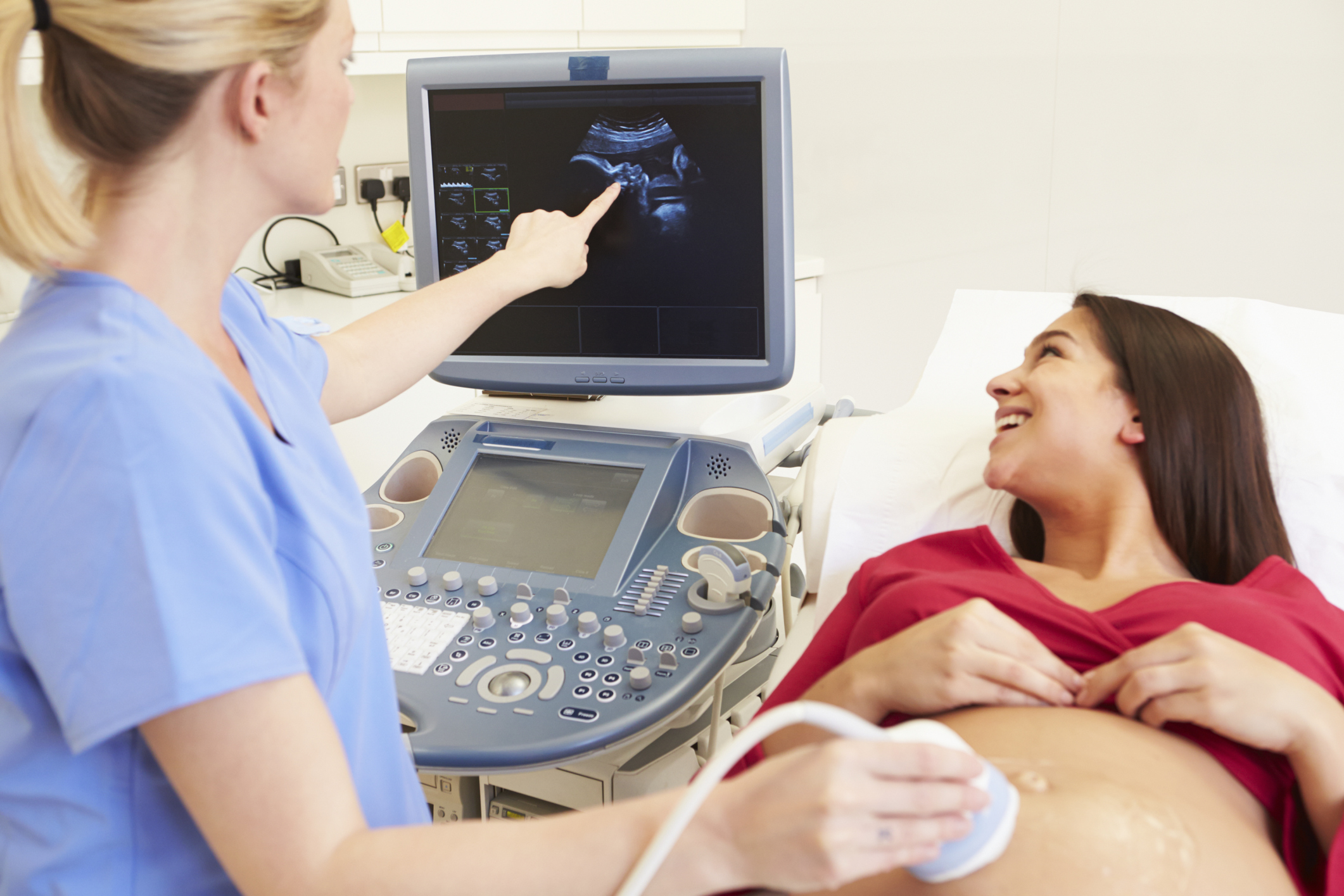
In an ultrasound, a legitimate wave is produced in short pulses on the desired frequency and targeted in the vicinity of interest inside the frame. These sound waves are in part contemplated returned from the frame, acquired by way of a transducer, and despatched to the ultrasonic scanner, in which they’re processed and converted into a digital photograph. The formation of the image depends on the time and the power of the echo and is displayed on the pc display screen for analysis.
CT experiment method
In the best diagnostic Center in Noida throughout CT scans, patients are moved through the scanning system. A supply of X-rays and an X-ray detector also rotate in synchronicity so that the digital X-rays which are handed through the region of interest can produce special image slices in axial or helical mode. The organized CT scan can be without delay viewed on a tv display or recorded for garage and evaluation later.
Ultrasound manner
In the course of clinical ultrasound, a probe is handed over to the location of interest to send sound waves into the area. To reduce air bubbles between the probe and the skin, a jelly is implemented in the area first. The patient is now and then asked to alternate positions to get a higher view of the target location. The pictures received measurements may be regarded or stored to be used later.
Programs of Ultrasound vs CT test
CT center in Noida has the benefits of CT scans are maximum glaring in screenings for cancer (tumors), accidents, or abnormalities in the body. The scans also can be combined with different strategies, along with ultrasound or Magnetic Resonance Imaging (MRI), for extra definition and precision.
Ultrasound is used for diagnostic programs, such as visualizing muscle tissues, tendons, and inner organs, to determine their size, structures, lesions, or different abnormalities. Obstetric sonography is used to visualize fetuses at some stage in pregnancy. Different applications of ultrasound include removing kidney and gall stones, lipectomy, and different packages. You can check online for the ultrasound center near me for the scanning process.

